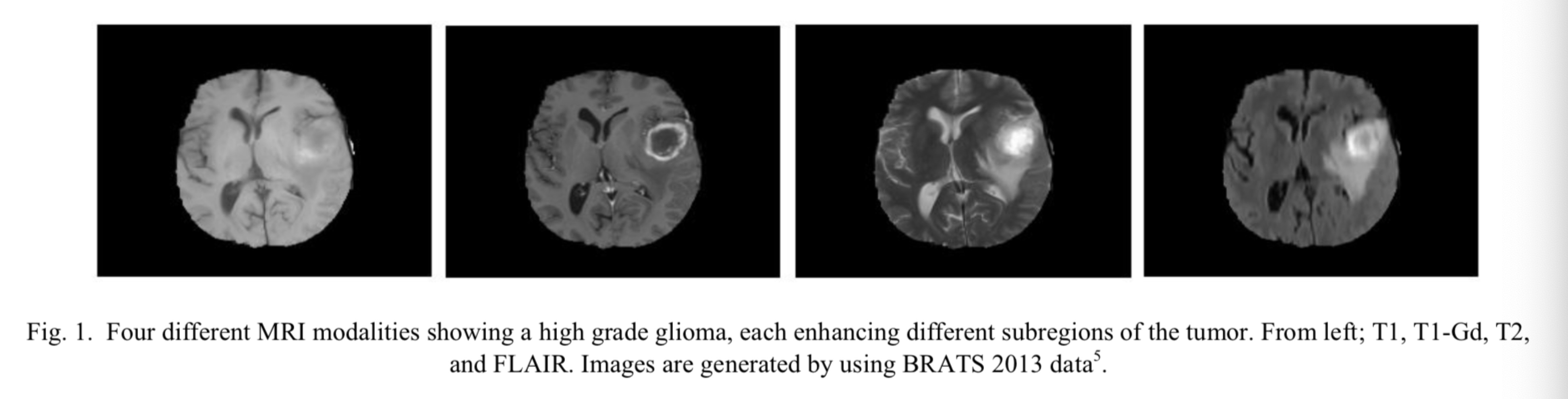
T1, T2, T1-Gd and FLAIR
13 Feb 2019T1: T1-weighted MRI
T2: T2-weighted MRI
T1-Gd: T1-weighted MRI with gadolinium contrast enhancement
FLAIR: Fluid Attenuated Inversion Recovery
T1 images are used for distinguishing healthy tissues, T2 images are used to delineate the edama rigion which produces bright signal on the image. T1-Gd images, the tumor border can easily be distinguished by the bright singal of the accumulated contrast agent(gadolinium ions) in the active cell region of the tumor tissue. Since necrotic cells do not interact with the contrast agent, they can be observed by hypo intense part of the tumor core making it possible to easily segment them from the active cell region on the same sequence. In FLAIR images, signal of water molecules are suppressed which helps in distinguishing edema region from the Cerebrospinal Fluid (CSF).